Link to our publications
-
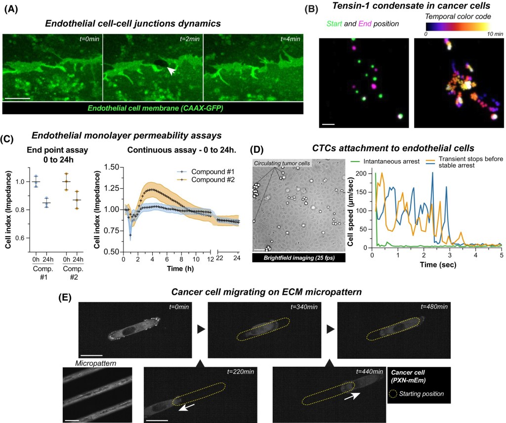
Fast label-free live imaging with FlowVision reveals key principles of cancer cell arrest on endothelial monolayers
The rapid, transient, and unpredictable nature of interactions between circulating cells and the endothelium challenges the investigation of these events under flow conditions. Here, we developed an imaging and image-analysis framework called FlowVision, which integrates fast, bright-field live-cell imaging with deep-learning-based image analysis to quantitatively track cell landing and arrest on an endothelial monolayer under…
-

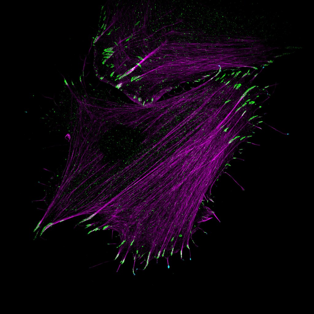
Filopodome proteomics identifies CCT8 as a MYO10 interactor critical for filopodia functions
Cancer cells utilize filopodia to explore, adhere to, and invade their surrounding microenvironment, yet the protein networks that organize these protrusions remain incompletely defined. To uncover the molecular machinery underlying MYO10-positive filopodia, we targeted the fast biotin ligase TurboID to the filopo-dia tip-localized motor protein MYO10. Proximity biotinylation in two cell types revealed hundreds of…
-

Customizable FDM-based zebrafish embryo mold for live imaging
Accurate and reproducible orientation of zebrafish embryos is crucial for high-resolution live imaging but remains challenging with standard agarose mounting. Here, we introduce an orientation tool made using fused deposition modeling (FDM), enhanced with a thin resin coating to improve surface smoothness and performance. This design creates reproducible, embryo-shaped wells that hold larvae in a…
-
Time, the final frontier
Cancer’s notorious heterogeneity poses significant challenges, as each tumor comprises a unique ecosystem. While single-cell and spatial transcriptomics advancements have transformed our understanding of spatial diversity within tumors, the temporal dimension remains underexplored. Tumors are dynamic entities that continuously evolve and adapt, and relying solely on static snapshots obscures the intricate interplay between cancer cells…
-
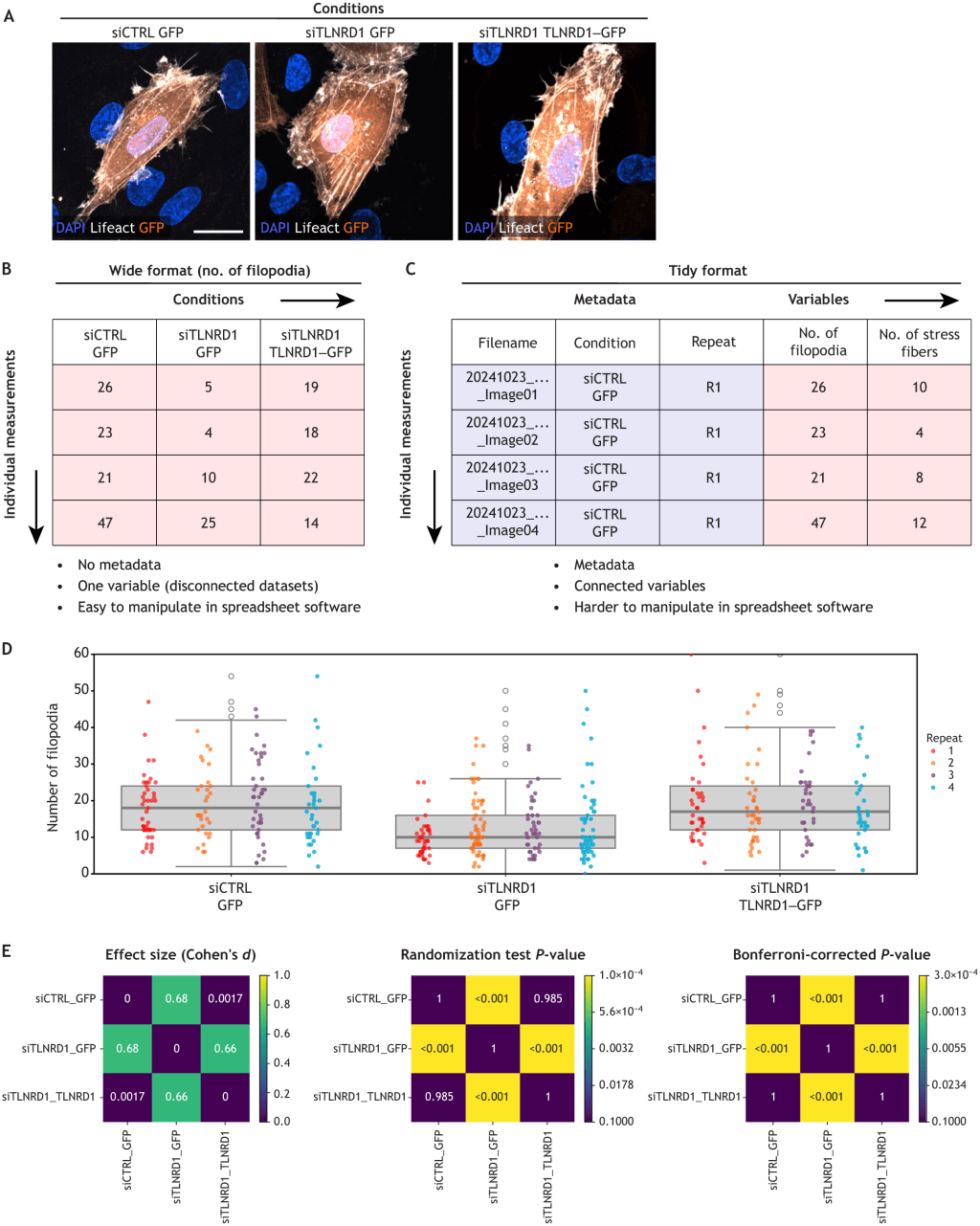
Practical considerations for data exploration in quantitative cell biology
Data exploration is an essential step in quantitative cell biology, bridging raw data and scientific insights. Unlike polished, published figures, effective data exploration requires a flexible, hands-on approach that reveals trends, identifies outliers and refines hypotheses. This Opinion offers simple, practical advice for building a structured data exploration workflow, drawing on the authors’ personal experience…
-
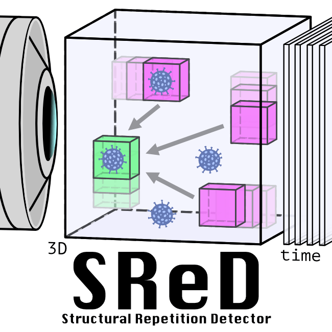
Structural Repetition Detector: multi-scale quantitative mapping of molecular complexes through microscopy
From molecules to organelles, cells exhibit recurring structural motifs across multiple scales. Understanding these structures provides insights into their functional roles. While super-resolution microscopy can visualise such patterns, manual detection in large datasets is challenging and biased. We present the Structural Repetition Detector (SReD), an unsupervised computational framework that identifies repetitive biological structures by exploiting…
-

PhotoFiTT: A Quantitative Framework for Assessing Phototoxicity in Live-Cell Microscopy Experiments
Phototoxicity in live-cell fluorescence microscopy can compromise experimental outcomes, yet quantitative methods to assess its impact remain limited. Here we present PhotoFiTT (Phototoxicity Fitness Time Trial), an integrated framework combining a standardised experimental protocol with advanced image analysis to quantify light-induced cellular stress in label-free settings. PhotoFiTT leverages machine learning and cell cycle dynamics to…
-

CellTracksColab is a platform that enables compilation, analysis, and exploration of cell tracking data
In life sciences, tracking objects from movies enables researchers to quantify the behavior of single particles, organelles, bacteria, cells, and even whole animals. While numerous tools now allow automated tracking from video, a significant challenge persists in compiling, analyzing, and exploring the large datasets generated by these approaches. Here, we introduce CellTracksColab, a platform tailored…
-

TLNRD1 is a CCM complex component and regulates endothelial barrier integrity
We previously identified talin rod domain-containing protein 1 (TLNRD1) as a potent actin-bundling protein in vitro. Here, we report that TLNRD1 is expressed in the vasculature in vivo. Its depletion leads to vascular abnormalities in vivo and modulation of endothelial cell monolayer integrity in vitro. We demonstrate that TLNRD1 is a component of the cerebral…
-

DL4MicEverywhere: Deep learning for microscopy made flexible, shareable, and reproducible
Iván Hidalgo-Cenalmor, Joanna W. Pylvänäinen, Mariana G. Ferreira, Craig T. Russell, Alon Saguy, Ignacio Arganda-Carreras, Yoav Shechtman, AI4Life Horizon Europe Program Consortium, Guillaume Jacquemet, Ricardo Henriques & Estibaliz Gómez-de-Mariscal Deep learning has revolutionised the analysis of extensive microscopy datasets, yet challenges persist in the widespread adoption of these techniques. Many lack access to training data, computing resources, and expertise to develop complex models. We introduce DL4MicEverywhere,…
-

Fast4DReg: Fast registration of 4D microscopy datasets
Unwanted sample drift is a common issue that plagues microscopy experiments, preventing accurate temporal visualization and quantification of biological processes. Although multiple methods and tools exist to correct images post-acquisition, performing drift correction of three-dimensional (3D) videos using open-source solutions remains challenging and time-consuming. Here, we present a new tool developed for ImageJ or Fiji…
-
MYO10-filopodia support basement membranes at pre-invasive tumor boundaries
Ductal carcinoma in situ (DCIS) is a pre-invasive stage of breast cancer. During invasion, the encapsulating DCIS basement membrane (BM) is compromised, and tumor cells invade the surrounding stroma. The mechanisms that regulate functional epithelial BMs in vivo are poorly understood. Myosin-X (MYO10) is a filopodia-inducing protein associated with metastasis and poor clinical outcome in…
-
Myosin-X recruits lamellipodin to filopodia tips
Myosin-X (MYO10), a molecular motor localizing to filopodia, is thought to transport various cargo to filopodia tips, modulating filopodia function. Yet, only a few MYO10 cargoes have been described. Here, using GFP-Trap and BioID approaches combined with mass spectrometry, we identified lamellipodin (RAPH1) as a novel MYO10 cargo. We report that the FERM domain of…
-

TrackMate 7: integrating state-of-the-art segmentation algorithms into tracking pipelines
TrackMate is an automated tracking software used to analyze bioimages and is distributed as a Fiji plugin. Here, we introduce a new version of TrackMate. TrackMate 7 is built to address the broad spectrum of modern challenges researchers face by integrating state-of-the-art segmentation algorithms into tracking pipelines. We illustrate qualitatively and quantitatively that these new…
-
TLNRD1 is a novel actin-bundling protein that promotes filopodia formation
Talin is a mechanosensitive adapter protein that couples integrins to the cytoskeleton. Talin rod domain–containing protein 1 (TLNRD1) shares 22% homology with the talin R7R8 rod domains and is highly conserved throughout vertebrate evolution, although little is known about its function. We show that TLNRD1 is an α-helical protein structurally homologous to talin R7R8. Like…
-
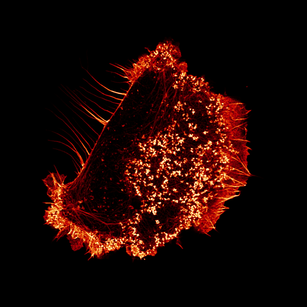
Myosin-X and talin modulate integrin activity at filopodia tips
Filopodia assemble unique integrin-adhesion complexes to sense the extracellular matrix. However, the mechanisms of integrin regulation in filopodia are poorly defined. Here, we report that active integrins accumulate at the tip of myosin-X (MYO10)-positive filopodia, while inactive integrins are uniformly distributed. We identify talin and MYO10 as the principal integrin activators in filopodia. In addition,…
-
Democratising deep learning for microscopy with ZeroCostDL4Mic
Deep Learning (DL) methods are powerful analytical tools for microscopy and can outperform conventional image processing pipelines. Despite the enthusiasm and innovations fuelled by DL technology, the need to access powerful and compatible resources to train DL networks leads to an accessibility barrier that novice users often find difficult to overcome. Here, we present ZeroCostDL4Mic,…
-

Automated cell tracking using StarDist and TrackMate
The ability of cells to migrate is a fundamental physiological process involved in embryonic development, tissue homeostasis, immune surveillance, and wound healing. Therefore, the mechanisms governing cellular locomotion have been under intense scrutiny over the last 50 years. One of the main tools of this scrutiny is live-cell quantitative imaging, where researchers image cells over…
-

Cell matrix adhesion in cell migration
The ability of cells to migrate is a fundamental physiological process involved in embryonic development, tissue homeostasis, immune surveillance and wound healing. In order for cells to migrate, they must interact with their environment using adhesion receptors, such as integrins, and form specialized adhesion complexes that mediate responses to different extracellular cues. In this review,…
-
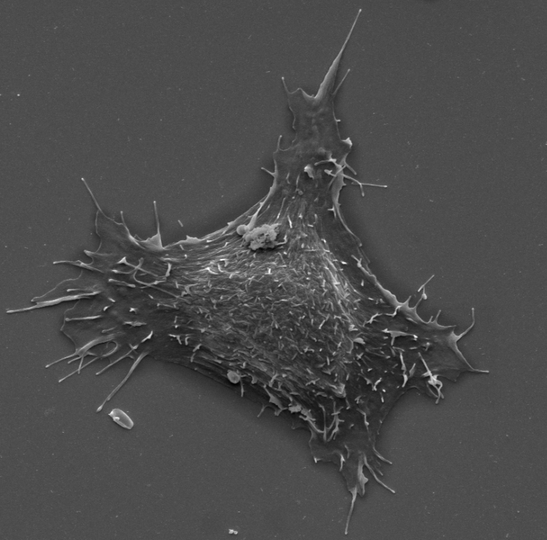
Filopodia in cell adhesion, 3D migration and cancer cell invasion
This review discusses recent advances in our understanding of the role filopodia and filopodia-like structures in cell adhesion and three dimensional (3D) cell migration both in vitro and in vivo. In particular, we focus on recent advances demonstrating that filopodia are involved in substrate tethering and environment sensing in vivo. We further discuss the emerging role of filopodia and…
-

Fluctuation-Based Super Resolution Traction Force Microscopy
Cellular mechanics play a crucial role in tissue homeostasis and are often misregulated in disease. Traction force microscopy is one of the key methods that has enabled researchers to study fundamental aspects of mechanobiology; however, traction force microscopy is limited by poor resolution. Here, we propose a simplified protocol and imaging strategy that enhances the…
