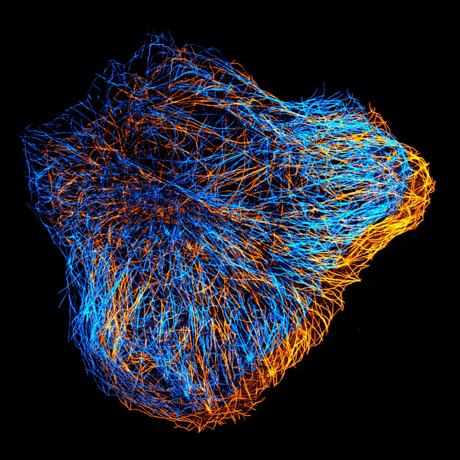
Cell biology, microscopy, and image analysis.
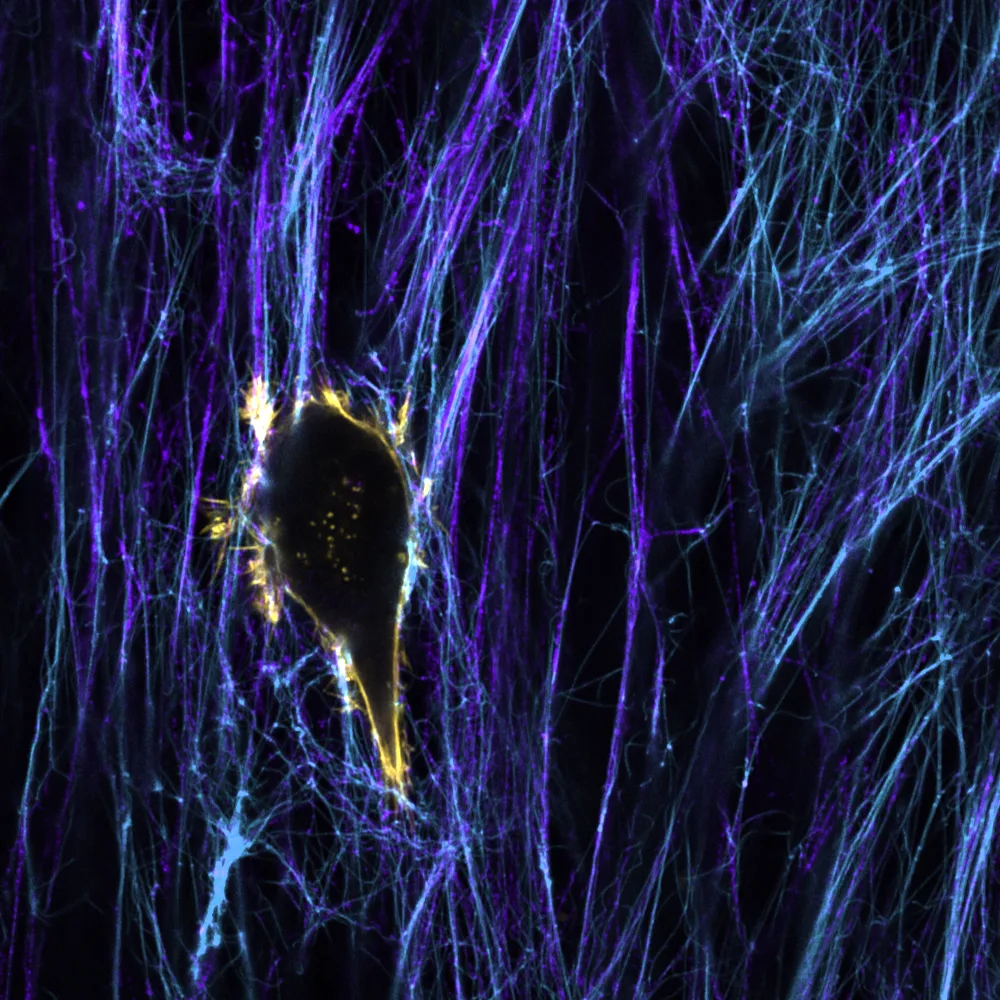
We study how cells move, interact with their surroundings, and cross tissue barriers. We combine cell biology with microscopy and quantitative image analysis, and we develop tools that make complex imaging experiments easier to analyse and share.
Featured research: biology
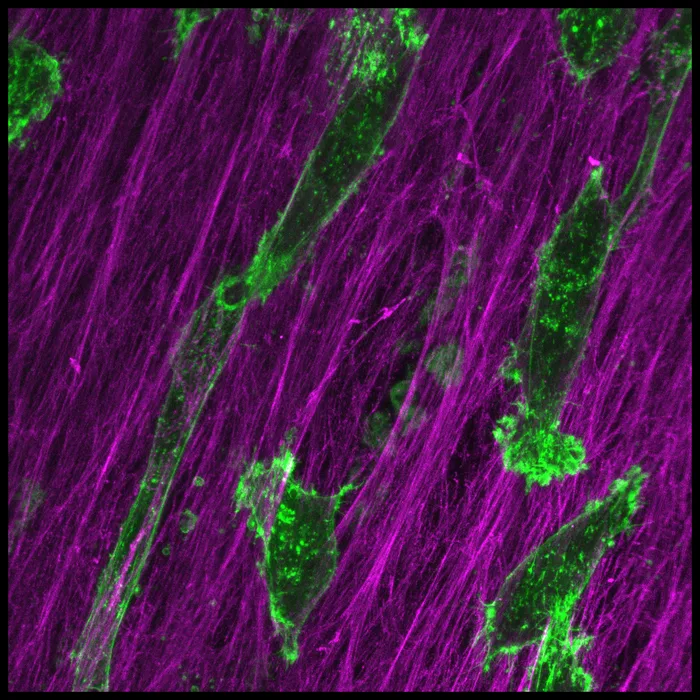
CCT8 associates with the MYO10 motor domain and regulates filopodia and breast cancer cell invasion
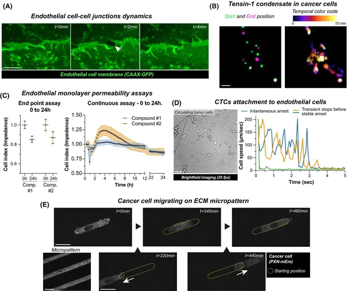
Pancreatic cancer cells breach endothelial barriers through protrusion-driven invasion or endothelial retraction
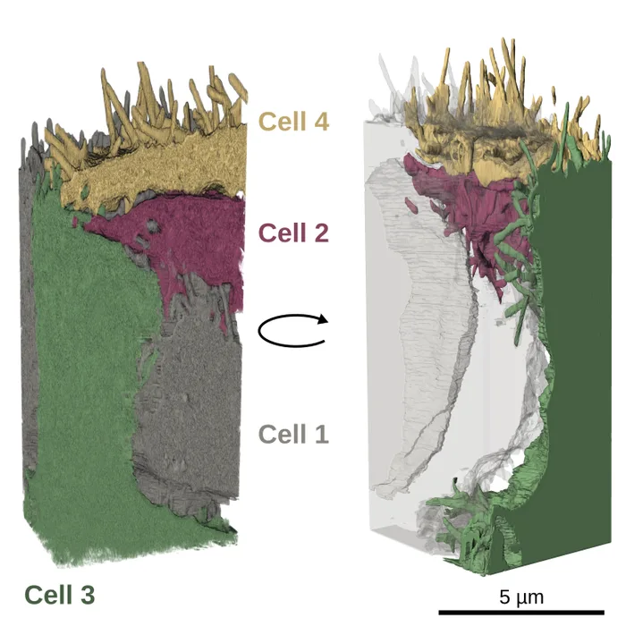
Filopodia-mediated trans-endocytosis
Fast label-free live imaging with FlowVision reveals key principles of cancer cell arrest on endothelial monolayers
Time, the final frontier
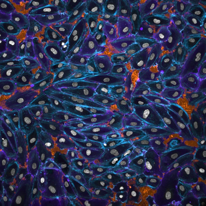
TLNRD1 is a CCM complex component and regulates endothelial barrier integrity
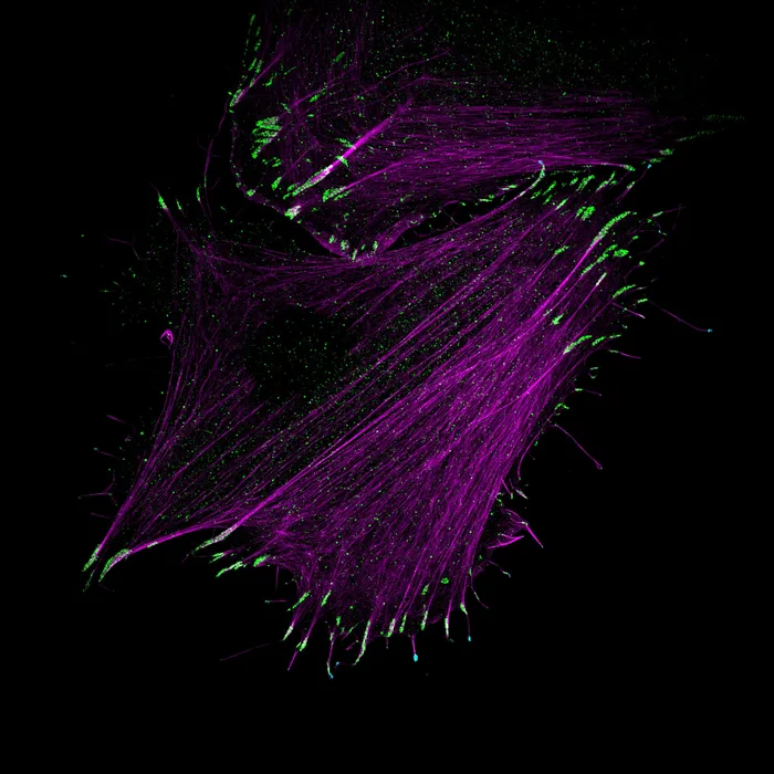
Myosin-X recruits lamellipodin to filopodia tips
MYO10-filopodia support basement membranes at pre-invasive tumor boundaries
Featured research: methods and tools

Rxiv-Maker – an automated template engine for streamlined scientific publications

NucleiSky enables cross-scale multimodal registration of microscopy data using nuclei constellations
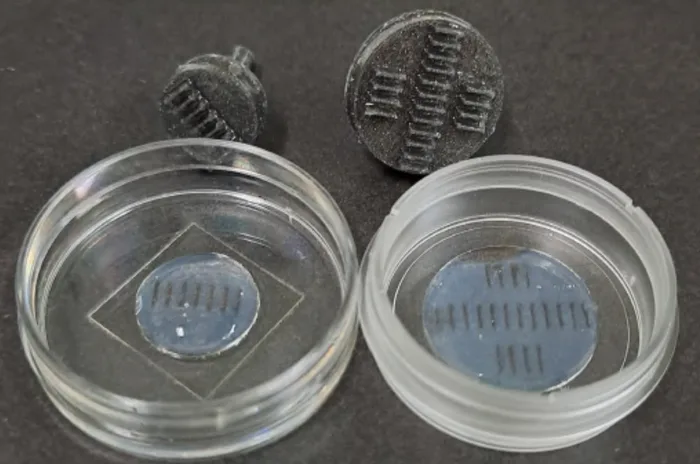
Customizable FDM-based zebrafish larvae mold for live imaging

Packaging Jupyter notebooks as installable desktop apps using LabConstrictor

EZInput: A Cross-Environment Python Library for Easy UI Generation in Scientific Computing
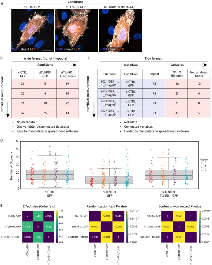
Practical considerations for data exploration in quantitative cell biology

CellTracksColab is a platform that enables compilation, analysis, and exploration of cell tracking data

DL4MicEverywhere: deep learning for microscopy made flexible, shareable and reproducible
All featured research (32) · All publications