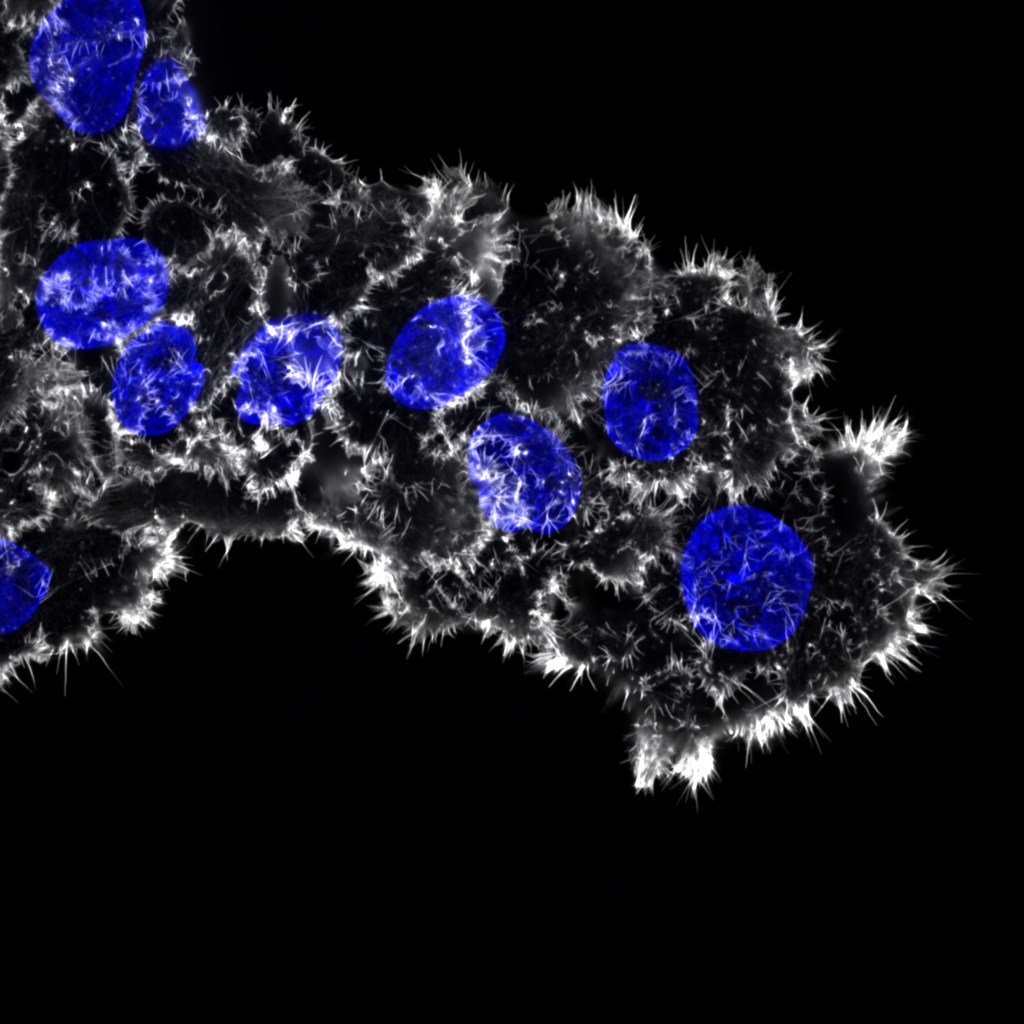
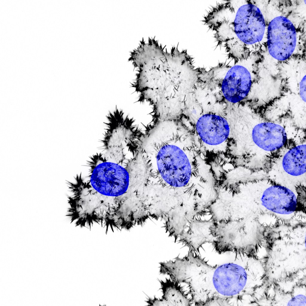
FiloQuant reveals increased filopodia density during breast cancer progression
Defective filopodia formation is linked to pathologies such as cancer, wherein actively protruding filopodia, at the invasive front, accompany cancer cell dissemination. Despite wide biological significance, delineating filopodia function in complex systems remains challenging and is particularly hindered by lack of compatible methods to quantify filopodia properties. Here, we present FiloQuant, a freely available ImageJ plugin, to detect filopodia-like protrusions in both fixed- and live-cell microscopy data. We demonstrate that FiloQuant can extract quantifiable information, including protrusion dynamics, density, and length, from multiple cell types and in a range of microenvironments. In cellular models of breast ductal carcinoma in situ, we reveal a link between filopodia formation at the cell–matrix interface, in collectively invading cells and 3D tumor spheroids, and the in vitro invasive capacity of the carcinoma. Finally, using intravital microscopy, we observe that tumor spheroids display filopodia in vivo, supporting a potential role for these protrusions during tumorigenesis.
Guillaume Jacquemet , Ilkka Paatero, Alexandre F. Carisey, Artur Padzik, Jordan S. Orange, Hellyeh Hamidi & Johanna Ivaska
Filopodia Quantification Using FiloQuant
Filopodia are fingerlike membrane protrusions that are extended by cells in vitro and in vivo. Due to important roles in sensing the extracellular microenvironment, filopodia and filopodia-like protrusions have been implicated in numerous biological processes including epithelial sheet zippering in development and wound healing and in cancer progression. Recently, there has been an explosion in the number of software available to analyze specific features of cell protrusions with the aim of gaining mechanistic insights into the action of filopodia and filopodia-like structures. In this methods chapter, we highlight an open-access software called FiloQuant that has been developed to specifically quantify the length, density, and dynamics of filopodia and filopodia-like structures from in vitro and in vivo generated samples. We provide step-by-step protocols on (i) how to install FiloQuant in the ImageJ platform (Fiji), (ii) how to quantify filopodia and filopodia-like protrusions from single images using FiloQuant, and (iii) how to track filopodial protrusions from live-cell imaging experiments using FiloQuant and TrackMate.
